Hippocampal Subfield Measurement and ILAE Hippocampal Sclerosis Subtype Classification with in Vivo 4.7 Tesla MRI.
March 1, 2020·,,,,,,·
0 min read
Trevor A. Steve
Justine Gargula
Ehsan Misaghi
Tomasz A. Nowacki
Laura M. Schmitt
B. Matt Wheatley
Donald W. Gross
Abstract
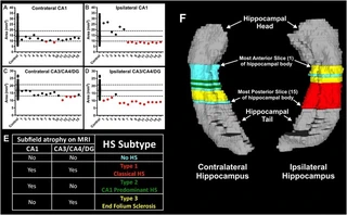
OBJECTIVE: Neuropathological studies indicate that hippocampal sclerosis (HS) consists of three subtypes (ILAE types 1-3 HS). However, HS subtypes currently can only be diagnosed by pathological analysis of hippocampal tissue resected during epilepsy surgery or at autopsy. In vivo diagnosis of HS subtypes holds potential to improve our understanding of these variants in the ipsilateral as well as contralateral hippocampus. In this study, we aimed to: i) evaluate the reliability of our histology-derived segmentation protocol when applied to in vivo MRI; and ii) characterize variability of HS subtypes along the hippocampal long axis in patients with epilepsy. METHODS: Eleven subjects with unilateral HS were compared with ten healthy controls. We used 4.7 T MRI to acquire high resolution MR Images of the hippocampus in each subject. In vivo MRI-based diagnoses of HS subtypes were then determined in each patient by two methods: i) hippocampal subfield volumetry of the entire hippocampal body; and ii) subfield area analysis at multiple thin slices throughout the hippocampal body. RESULTS: Hippocampal body subfield segmentation demonstrated excellent reliability and volumetry of the symptomatic hippocampus revealed abnormalities in all eleven patients. Six subjects demonstrated findings consistent with type 1 HS while five subjects had volumetry-defined atypical HS (two with type 2 HS & three with type 3 HS) in the symptomatic hippocampus, while five subjects were found to have type 3 HS in the contralateral hippocampus. Subfield area analyses demonstrated remarkable variability of HS subtypes along the hippocampal long axis, both ipsilateral and contralateral to the seizure focus. SIGNIFICANCE: Our results provide preliminary evidence that determining HS Subtype using in vivo MRI may allow preoperative diagnosis of ILAE HS subtypes. Further studies are essential to determine the pathological correlates of these neuroimaging findings. The heterogeneity of abnormalities observed along the long axis of the hippocampus is consistent with previous autopsy studies and highlights the necessity of studying the entire hippocampus both ipsilateral and contralateral to the seizure focus in these future studies.
Type
Publication
Epilepsy research
Adult
Epilepsy Temporal Lobe
Epilepsy Temporal Lobe/Pathology/*Surgery
Female
Hippocampal Sclerosis (HS)
Hippocampal Subfields
Hippocampus
Hippocampus/*Pathology
Humans
Image Processing Computer-Assisted
Image Processing Computer-Assisted/Methods
In Vivo Magnetic Resonance Imaging (MRI)
Magnetic Resonance Imaging
Magnetic Resonance Imaging/Methods
Male
Middle Aged
Reproducibility of Results
Sclerosis
Sclerosis/*Pathology
Seizures
Seizures/*Pathology
Temporal Lobe Epilepsy (TLE)
Young Adult
Authors
Authors
Authors
Authors
Authors
Authors
Authors